Looking for information about other Dental Topics?
Full Website Index• Animated-Teeth.com •

How many roots and root canals does a tooth have?
How many roots does each tooth have? | How many root canals are in each root?
This guide provides an overview of the typical root and root canal configuration of each kind of tooth (molar, premolar, canine, and incisor). It also explains how dentists identify a tooth’s root canals using methods like X-rays, visual inspection, and tactile exploration.
By understanding these details, patients can better appreciate the steps involved in performing their endodontic therapy and the difficulty a tooth’s anatomy sometimes presents in addressing all parts of its root canal system.
How many roots and root canals do teeth have?
(And why are these numbers important to know.)
When endodontic therapy is performed for a tooth, the number of roots and root canals being treated is an important factor because so many issues associated with this procedure are influenced by the total number of both. For example:
- The level of difficulty associated with performing a tooth’s work will depend on its number of roots, number of canals, and how complex its combined root canal configuration is.
- The success of endodontic therapy can depend on how accurately the dentist interprets the tooth’s root and root canal morphology. The same goes for avoiding potential complications during a tooth’s treatment.
- How long or how many visits What’s common? a tooth’s procedure will take depends on the number of roots and canals being treated.
- Even how much a tooth’s treatment will cost Endodontic fees by tooth type. is influenced by its number of roots and canals.
Variation is commonplace and must be expected.
What’s usually found vs. what is actually found.
In our chart below you’ll find the tally of what your dentist generally expects to discover when treating different types of teeth (molars, premolars [bicuspids], canines [cuspids], and incisors). However, encountering variation is a key theme when it comes to these numbers.
How many roots and canals your dentist actually finds, and therefore must ultimately treat when performing its endodontic therapy, can vary, even substantially, from what is considered “normal.”
In fact, the only valid axiom that can be given about this subject is that every tooth has at least one root and each root always contains at least one canal. In practice, the canal may be very tiny and difficult to locate. But a dentist always knows to look for at least one root canal in each tooth root.
▲ Section references – Torabinejad
So, how many roots and root canals does your tooth have?
This answer will primarily depend on which type of tooth is being considered. Here are the “normal” numbers for different kinds of teeth.
Number of roots - Permanent / Adult teeth.
- How many roots does an incisor have (upper or lower)? – One.
- How many roots does a canine have (upper or lower)? – One.
- How many roots does an upper premolar have? – One or two.
- How many roots does a lower premolar have? – One.
- How many roots does an upper molar have? – Three.
- How many roots does a lower molar have? – Two.
Number of root canals - Permanent / Adult teeth.
- How many root canals does an incisor have (upper or lower)? – One.
- How many root canals does a canine have (upper or lower)? – One.
- How many root canals does an upper premolar have? – Two.
- How many root canals does a lower premolar have? – One.
- How many root canals does an upper molar have? – Three or more.
- How many root canals does a lower molar have? – Three or more.
Here’s the same information in tabular form.
Table – Usual root and root canal expectations for Molars, Premolars, Canines, and Incisors. - Adult / Permanent teeth.
| Kind of tooth. | How many roots? | How many root canals? |
|---|---|---|
| Upper Incisors Central Incisors Lateral Incisors | 1 | 1 |
| Upper Canines | 1 | 1 |
| Upper 1st Premolars | 1 or 2 | 2 |
| Upper 2nd Premolars | 1 | 1 or 2 |
| Upper Molars First Molars Second Molars Third Molars (Wisdom Teeth) | 3 | 3 or more |
| Lower Incisors Central Incisors Lateral Incisors | 1 | 1 |
| Lower Canines | 1 | 1 |
| Lower Premolars First Premolars Second Premolars | 1 | 1 |
| Lower Molars First Molars Second Molars Third Molars (Wisdom Teeth) | 2 | 3 or more |
Your dentist must assume that every tooth displays variation in its number of canals and possibly roots too.
As stated above, while the numbers shown in our chart are the common and usual ones for each kind of tooth, the bottom line is that when performing any one tooth’s endodontic work the dentist must look for what’s expected, and then anticipate that they’ll find more. This definitely goes for the total number of root canals in each root and possibly even the total number of roots the tooth has too.
This is vital because a tooth’s root canal therapy won’t be successful unless the dentist treats the tooth’s entire root canal system. Any portion that’s overlooked Missed canals., and therefore is not treated, can be expected to result in treatment failure.
Forms of anatomical variation your dentist is likely to discover.
- Additional tooth roots – While it’s always possible that your tooth has a greater number of roots than most other teeth of its type, as compared to variations in the number of root canals, this type of deviation is less common.
A primary exception to this rule would be upper premolars (bicuspids) where statistically speaking the presence of either just one or two roots runs practically neck and neck. (Al-Ghananeem, Chaparro)
- Fused tooth roots – More common than finding extra roots is the issue of multi-rooted teeth (premolars, molars) having fused roots.
This issue on its own doesn’t necessarily increase the difficulty of a root canal case. But it’s not uncommon that teeth with fused roots display wider variation in their number of root canals and the complexity of their configuration, which would tend to do so. (Ahmad)
- Additional root canals – Discovering a tooth that has a greater number of canals than what’s listed in our table above isn’t uncommon at all.
Certain types of teeth (lower incisors, upper premolars, molars) and even specific tooth roots (mesiobuccal roots of upper first molars, distal roots of lower 1st molars) are well known for having the potential to have additional canals.
▲ Section references – Al-Ghananeem, Chaparro, Ahmad
How does a dentist identify and locate all of a tooth’s roots and root canals?
If variation in the number of roots and canals is so common and so important for the dentist performing the tooth’s endodontic treatment to know about, how do they discover them all?
The three primary methods clinicians use are:
a) Taking X-rays.
Long before your dentist ever begins performing your tooth’s actual root canal work, they’ll have taken dental X-rays. And these pictures of your jaw can give them a substantial amount of information about how many roots and root canals your tooth has.
1) Two-dimensional pictures.
The most common type of dental X-ray general dentists take is like the one shown in our graphic. It’s a flat (two-dimensional) picture of your three-dimensional tooth.
What your dentist can learn from this type of film.
An X-ray like the one shown below can reveal a lot of information. Here are some of the details a dentist would notice: (The tooth in our picture is a lower first molar.)
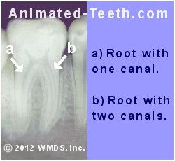
- Running down the length of root “B” you can see the clear outline of two root canals.
Actually, that’s to be expected. This root (the mesial root) of a lower 1st molar usually does have two canals.
- In contrast to root “B,” when you look at root “A” you just see one canal. And actually, for this root (the distal root) that’s the most common form.
- We’ll also point out that from this picture it’s clear that this tooth has two distinct roots (as opposed to them being fused together), which once again is the most common form for this type of tooth.
So, just from the simple act of taking a radiograph, and long before they have taken any instruments to your tooth at all, it’s possible for your dentist to have quite a bit of information about it. Both in terms of how many roots and canals your tooth has. And with this particular tooth in our example, so far everything seems usual about it.
What your dentist can’t always tell from a two-dimensional X-ray.
As informative as taking conventional dental X-rays can be, there can be times when what they show can be difficult to interpret. To give you some insight into what your dentist must deal with, here are some unclear points about the same radiograph previously shown above.
Do root “B”‘s canals merge?
Root “B”‘s two separate canals are obvious at the level of the arrow. But notice how down around the tooth’s tip they aren’t so distinct?
What’s the real story? Are these two full-length canals, or do they possibly merge into a single one down low?
It’s most likely that due to the angle at which this film was taken, one canal simply overlaps the other (the two are superimposed) thus giving the appearance of just one.
Why dentists take multiple X-rays.
Actually, your dentist has a technique they can use that can help to clarify things. That’s simply taking an additional X-ray(s) from a slightly different angle (like plus or minus 20 degrees) and comparing them.
When that’s done, what actually exists is more likely to be revealed. And for this reason, it’s commonplace and should be expected that your dentist will take multiple (two or more) pre-operative X-rays of your tooth.
Does root “A” have one or two canals?
This same type of inconclusiveness exists with root “A”. It looks like it just has a single canal but it’s not uncommon for a first molar’s distal root to have two. (About one-third of cases have two. Ingle – linked above.)
Once again, the angle at which this picture was taken may have resulted in superimposing the two canals. And also just like above, taking additional radiographs from different angles may help to clarify what actually exists.
2) Three-dimensional radiographs.
Over the past decade, the use of Cone Beam Computed Tomography (CBCT) 3D imaging. has become more and more common in dentistry. And kinds of images that this technique can provide can go a long way in answering uncertainties about a tooth’s roots and canals, especially for teeth that have an abnormal or very complex anatomy.
Due to the expense of these units, however, most general practitioners don’t have CBCT capability. Its use with root canal work is typically limited to the offices of root canal specialists Endodontists.
b) Searching for root canals via visual inspection.
Once your dentist has begun the process of performing your tooth’s endodontic therapy, visual inspection is a vitally important way by which they determine how many root canals it has.
The picture below shows the inside of a lower first molar (the same type of tooth as in our example above). This is what the dentist sees after creating the opening access cavity through which they’ll perform their work.
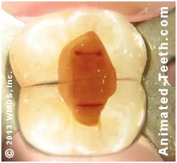
The pulpal floor of a lower first molar.
- What you see is the floor of the tooth’s pulp chamber (its “pulpal floor”).
- The openings you see (the two faint lines inside the tooth) are literally the openings (orifices) of each of the tooth’s root canals where they connect with the tooth’s pulp chamber.
When examining this picture, a dentist would notice:
- Just like described above, the mesial root (which lies underneath the portion of the tooth at the top of our picture) appears to have two separate canals. (You can see the two small round openings at each end of the faint dark line).
- And just like above but in contrast to the mesial root, the distal root (which lies underneath the portion of the tooth at the bottom of our picture) seems to have just one broad slit-like canal.
The dentist will need to investigate further to confirm that this is in fact just a single canal.
Inspection via microscope.
Visual inspection is such an important discovery tool that it’s common for root canal specialists to use a surgical microscope when they evaluate a tooth’s pulpal floor.

An endodontist using an operating microscope.
An example from research.
Initially, 73% of teeth were identified as having a second canal in this root. However, with the aid of a microscope, it was determined that a second canal was actually present in 93% of cases.
This type of discovery is important because missing (not treating) a canal, even one this small, can be expected to lead to root canal failure. In fact, a study by Hoen suggests that 42% of failed cases involve missed canals.
▲ Section references – Stropko, Hoen
c) Tactile discovery of root canals during your procedure.
Another important way by which a tooth’s total number of root canals is ultimately ascertained is by tactile discovery during the process of performing its endodontic work.
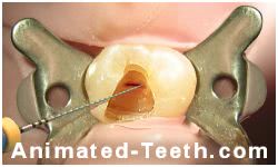
Working a file inside a tooth’s root canal.
Dentists clean and shape the nerve space The steps. inside a tooth via the use of root canal files. And the paths these files tend to take as they are worked up and down inside the tooth’s root canal system give the dentist a good idea of its overall anatomy.
Configurations your dentist may discover.
It’s possible for a dentist to ascertain various factors about a tooth’s canal system as they work.
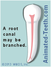
- The canal may be branched. – A single canal sometimes divides into two separate ones.
With our distal root example above, that was the concern. And as the dentist performs the tooth’s work, they would need to investigate whether what looks like the opening to a single canal is in fact that. Or if instead, it branches into two separate ones lower on down in the root.
- Two canals may coalesce into one. – We mentioned the possibility of this configuration in our mesial root example above. It’s possible that two obviously separate canals at the tooth’s pulpal floor combine to form a single one lower on down in the root.
Now with the kind of tooth in our example above that’s unlikely. But the dentist must confirm this fact as they perform their work.
- The issue of how many roots a tooth has does vary for some kinds of teeth. But with others, variation is so rare that it’s practically a nonissue.
- In comparison, how many root canals a tooth has tends to vary much more frequently. This variation will be more common with specific kinds of teeth and specific tooth roots on certain types of teeth.
- And because discovering and treating all canals is so vitally important for the success of a tooth’s endodontic work, dentists place great emphasis on searching for and identifying them using the methods described on this page.
What’s next?
We have a lot more information about root canal treatment …
Last reviewed: January 10, 2025
Author: Paul Cotner, DMD — retired dentist.
Published by: WMDS, Inc. — owner of Animated-Teeth.com.
Educational information only — not a substitute for professional dental care.
Page references sources:
Ahmad IA, et al. Root and root canal morphology of third molars in a Jordanian subpopulation.
Ingle JI, et al. Ingle’s Endodontics. Chapter: Morphology of Teeth and Their Root Canal Systems.
Al-Ghananeem MMF, et al. The Number of Roots and Canals in the Maxillary Second Premolars in a Group of Jordanian Population.
Chaparro AJ, et al. Number of roots and canals in maxillary first premolars: study of an Andalusian population.
Hoen MM, et al. Contemporary Endodontic Retreatments: An Analysis based on Clinical Treatment Findings.
Stropko JJ. Canal morphology of maxillary molars: clinical observations of canal configurations.
Torabinejad M, et al. Endodontics. Principles and Practice. Chapter: Endodontic radiography.