Looking for information about other Dental Topics?
Full Website Index• Animated-Teeth.com •

Canker Sores vs. Herpes – What’s the difference between these kinds of oral lesions?
Guide to differentiating herpetic and aphthous (canker) oral lesions.
Canker sores and oral herpes are common oral lesions that are often confused. However, they differ significantly in causes, symptoms, appearance, and treatment.
This guide offers a comprehensive comparison between canker sores and oral herpes lesions, highlighting their distinguishing features to help you accurately identify and understand each condition. (Check out this page’s Table of Contents for a complete list of its topics.)
Herpetic and Aphthous oral lesions are often confused.
As a start for this guide, we need to sort through a few different comparisons.
a) Canker sores vs. Oral herpes.
Of all of the different kinds of mouth ulcers that are commonly mistaken for canker sores (more formally known as recurrent minor aphthous ulcers), the one most often confused in identification is the recurrent intraoral herpes lesion.
The term “intraoral” indicates that this form of herpes lesions occur inside the mouth, just like canker sores. And the focus of this page is how to tell these two kinds of mouth ulcers apart.
b) Canker sores vs. Cold sores.
As opposed to intraoral herpetic lesions, another form of sore caused by the herpes virus is cold sores (also known as fever blisters).
Since these lesions form outside the mouth (on the lips or on nearby facial skin) it’s fairly impossible to mistake them for canker sores. (FYI: If you’d like more information about Cold Sore Identification use this link.)
c) Are canker sores caused by herpes or any other kind of virus?
No, they are not. Canker sores are caused by an inappropriate response of a person’s own immune system, not a virus.
While this page focuses on lesion diferentiation, these links may be of interest to you as further information about the causes of canker vs. herpetic sores.
- The biological mechanism (immune system response) that causes canker sores to form.
- Triggers for canker sore outbreaks.
- Herpes virus infection and the types of lesions that it can cause.
Canker Sore or Herpes? quiz – A quick way to learn the important points from this page.
https://www.animated-teeth.com/?p=182#quiz
Canker sores vs. Oral herpes – How can you tell the two apart?
There are some very distinct differences between herpes and aphthous lesions, and they are very easy to identify. The remainder of this page explains what to look for.
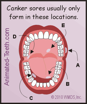
A) Canker sores and oral herpes form in different locations.
Compare: Intraoral herpes
Herpes lesions form on keratinized oral tissues. These are the “tougher,” firmer type of gum tissue found in the mouth.
Keratinized tissues are sometimes described as being bone-bearing, meaning that they’re tightly bound as a covering over the bony structures that lie underneath them.
Keratinized oral tissues include: (See Frame 1 of our animated graphic.)
a) The skin covering the hard palate. – This is the most likely location for intraoral herpetic lesions to form.
b) Attached gingiva. – This term refers to the gum tissue that surrounds the teeth and covers over the portion of the jawbone that encases them.
Canker sores vs. Oral herpes.
Oral herpes (Slide 1) and canker sores (Slide 2) form in different locations.
Compare: Canker sores
- a) The inside lining of the cheeks or lips.
- b) The underside of the tongue.
- c) The floor of the mouth.
- d) In the back of the mouth, near the tonsils.
- e) The soft palate.
▲ Section references – Neville, Scully, Mortazavi, Dunlap
FYI – You should be able to base most of your differentiation process just on the location of the sore alone. There can be exceptions but not usually with the type of common run-of-the-mill lesions you’re most likely to encounter.
Also note that different than with both aphthous ulcers and intraoral herpetic lesions, cold sores (also caused by the herpes simplex virus) form external to the mouth (on the lips and nearby facial skin). Differentiating cold sores from canker sores and intraoral herpes is a no-brainer just using location alone.
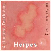
B) The early stages of canker and herpes sores look different.
Compare: Intraoral herpes
Herpetic sores first appear as a group of tiny vesicles (blisters).
- Each one is usually no more than about 1/25 of an inch (1mm) across.
- In our animated graphic below, you can actually see a vesicle in the lower right corner that hasn’t yet burst.
Compare: Canker sores
- These lesions first appear as a reddened area (a macule) on the surface of the mouth’s soft tissue lining and frequently cause a tingling, itching, or burning sensation.
- The area then transforms into an ulcer that may become as large as 1/4 of an inch.
FYI – Blister formation is a primary key in differentiating between aphthous and herpetic lesions.
- If you see tiny blisters form, you don’t have a canker sore. However, if you don’t see them it doesn’t absolutely rule out herpes.
- That’s because after herpetic blisters form they tend to burst fairly quickly, possibly before you’ve had a chance to actually notice them.
Oral herpes vs. Canker sores.
The shape of intraoral herpes (Slide 1) is entirely different than a canker sore (Slide 2).
C) The shape of the ulcerations is different.
Compare: Intraoral herpes
Compare: Canker sores
With aphthous lesions, the outline of the ulcerated area is usually round or ovoid and is surrounded by a smooth, regular (not jagged) red border. (See Frame 2 of our animated graphic.)
FYI – When shape is used to distinguish between the two, the rule of thumb to use is:
- Round or ovoid lesions = canker sores.
- Irregular shapes = oral herpes.
D) Distribution in the mouth.
In the case where you’ve had the same type of mouth sore before, the location where your current one has formed can help you to make a distinction between the two types of lesions.
- Oral herpes tends to recur in the same general area as before.
- In comparison, aphthous ulcers don’t necessarily show this same correlation. They often appear in entirely different areas each time.
FYI – When location is used to distinguish between the two, the rule of thumb to use is:
- If the location is always pretty much the same = oral herpes.
- If it’s more variable = canker sores.
E) Accompanying symptoms outside the mouth.
Compare: Intraoral herpes
With herpes, a person may experience malaise (bodily weakness or discomfort), fever, joint pain, or swollen lymph nodes in the neck. Although, these symptoms may be slight enough that you don’t really notice them.
Compare: Canker sores
None of the above symptoms are typical. In fact, the most common set of events is one where a lesion has formed but otherwise you feel completely normal.
FYI – If you’ve been sick, or even just a little under the weather, think herpes outbreak.
F) Other tip-offs.
The formation of a canker sore is sometimes preceded by some type of traumatic act, like biting or scraping the soft lining of your mouth.
In comparison, intraoral herpes lesions are often triggered by some type of traumatic event, with dental work frequently being the cause. As examples:
- Receiving a dental injection (getting a “shot”).
- Having some type of periodontal (gum) procedure performed in the area of the breakout.
FYI – The majority of reference sources used to create this page (see links above) specifically cite the above two dental procedures as the classic triggering events for intraoral herpes.
G) Healing time frames.
Both types of ulcerations can be expected to heal on their own within a 1 to 2 week time period.
Persistent sores require evaluation.
Mouth sores that persist for longer than two weeks may still be canker or intraoral herpes lesions. But this delay suggests that complicating factors are involved and closer evaluation is needed so appropriate treatment can be started.
- The delay might be due to some persistent source of mechanical irritation (like a broken tooth or sharp denture edge).
- Biopsy evaluation may be needed so the lesion can be differentiated from other types of mouth sores (including squamous cell carcinoma).
- With herpes, people having a compromised immune system (such as that due to HIV/AIDS, leukemia, or taking organ-transplant medications) may experience persistent ulcerations (that may even spread). In these cases, the use of antiviral drugs is likely indicated.
- Persistent canker ulcerations, or frequent or severe outbreaks, can be a symptom of an undiagnosed underlying medical condition See the list.. Or, the lesion may be a different form of aphthous ulcer (major aphthae How they differ.).
FYI – If the course of the ulceration you’ve been monitoring begins to stray from the expected norm, it requires evaluation by your dentist.
The hope is that the problem they discover is easily resolved. For example, possibly a rough tooth or denture flange just needs to be polished down.
But in some instances, they may find a serious situation that without treatment might have led to a severely extended or complicated healing process, or possibly even a life-threatening event.
▲ Section references – Primary reference sources for this entire page: Neville, Scully, Mortazavi, Dunlap
H) Contagiousness.
While not a factor that you would hope would be one by which these two types of mouth sores are differentiated, herpetic lesions are contagious whereas canker sores are not.
Compare: Intraoral herpes
The causative infectious agent for herpetic lesions is the herpes simplex virus (HSV-1 or HSV-2). And it proliferates within the sores that it forms.
Therefore, due to the potential for viral transmission from lesions, intraoral herpes is contagious. Exposure to other’s lesions, even via an intermediary object (like an eating or drinking utensil) can spread the disease.
Compare: Canker sore.
Canker sore formation is due to the response of your own body’s immune system to the presence of antigens (foreign molecules). There’s no infectious agent to spread. Therefore, canker sores are not contagious.
More details: Why canker sores aren’t contagious. The biology of canker sore formation.
FYI – If someone you’ve been in close contact with during the period when you’ve had sores then later on develops the same type of lesions for the first time, think herpes.
Looking for more information?
If you’re looking for additional information about canker sores, then we would suggest choosing from this menu: Canker Sore Guides
Use this link if you’d like more information specifically about intraoral herpetic lesions.
The herpes virus also causes cold sores (a type of extraoral lesion). Here’s our offering of pages that cover them: Cold Sore/Fever Blister Guides
Last reviewed: April 25, 2026
Author: Paul Cotner, DMD — retired dentist.
Published by: WMDS, Inc. — owner of Animated-Teeth.com.
Educational information only — not a substitute for professional dental care.
Page references sources:
Dunlap CL, et al. A guide to common oral lesions.
Mortazavi H, et al. Diagnostic Features of Common Oral Ulcerative Lesions: An Updated Decision Tree.
Neville BW, et al. Oral and Maxillofacial Pathology. Chapter: Allergies and Immunologic Diseases.
Scully C, et al. Oral medicine — Update for the dental practitioner Aphthous and other common ulcers.